Photoacoustic imaging combines optical and ultrasound technologies to provide high-contrast, high-resolution images by detecting ultrasonic waves generated from laser-induced thermoelastic expansion in tissues. Unlike traditional ultrasound imaging, which relies solely on acoustic impedance differences, photoacoustic imaging offers enhanced molecular and functional information, enabling better visualization of blood oxygenation and tumor angiogenesis. This hybrid technique improves early disease diagnosis and monitoring by integrating structural and biochemical insights in a single modality.
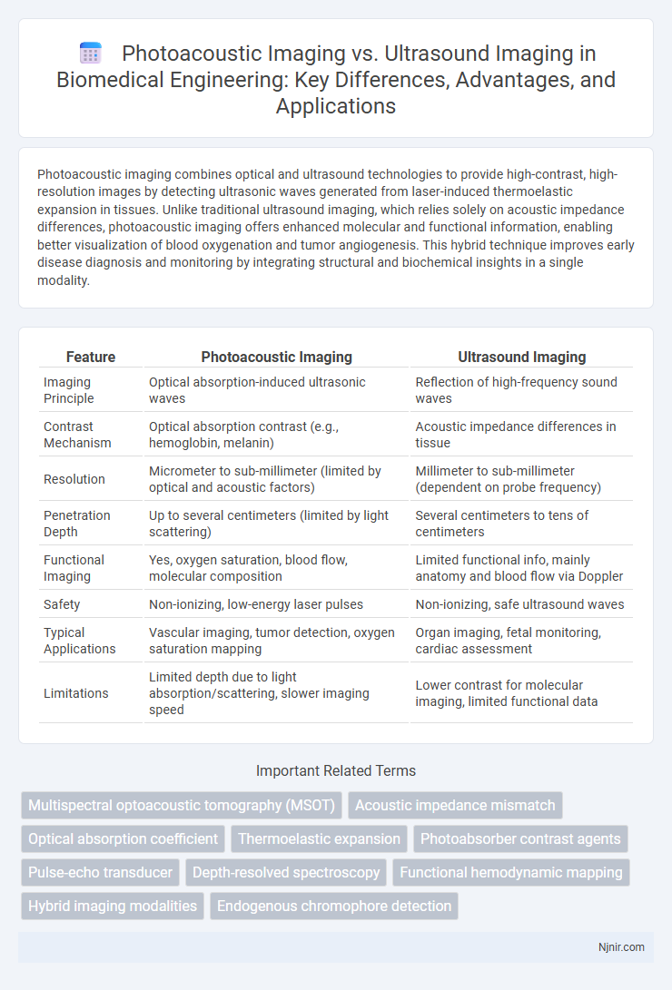
Table of Comparison
| Feature | Photoacoustic Imaging | Ultrasound Imaging |
|---|---|---|
| Imaging Principle | Optical absorption-induced ultrasonic waves | Reflection of high-frequency sound waves |
| Contrast Mechanism | Optical absorption contrast (e.g., hemoglobin, melanin) | Acoustic impedance differences in tissue |
| Resolution | Micrometer to sub-millimeter (limited by optical and acoustic factors) | Millimeter to sub-millimeter (dependent on probe frequency) |
| Penetration Depth | Up to several centimeters (limited by light scattering) | Several centimeters to tens of centimeters |
| Functional Imaging | Yes, oxygen saturation, blood flow, molecular composition | Limited functional info, mainly anatomy and blood flow via Doppler |
| Safety | Non-ionizing, low-energy laser pulses | Non-ionizing, safe ultrasound waves |
| Typical Applications | Vascular imaging, tumor detection, oxygen saturation mapping | Organ imaging, fetal monitoring, cardiac assessment |
| Limitations | Limited depth due to light absorption/scattering, slower imaging speed | Lower contrast for molecular imaging, limited functional data |
Introduction to Biomedical Imaging Modalities
Photoacoustic imaging combines optical absorption contrast with ultrasonic spatial resolution, enabling visualization of vascular structures and oxygen saturation in tissues. Ultrasound imaging uses high-frequency sound waves to produce real-time images of soft tissues, widely applied for anatomical and functional assessment. Both modalities are essential in biomedical imaging, with photoacoustic imaging offering molecular-level information and ultrasound providing dynamic structural insights.
Principles of Photoacoustic Imaging
Photoacoustic imaging operates by converting absorbed optical energy into ultrasonic waves through the photoacoustic effect, where pulsed laser light induces localized thermoelastic expansion in tissues. Unlike conventional ultrasound imaging that relies solely on sound wave reflection, photoacoustic imaging combines optical contrast with ultrasonic detection, enabling visualization of functional and molecular information in biological tissues. This principle allows for high-resolution imaging with enhanced contrast based on optical absorption properties, particularly useful for vascular and oxygenation mapping.
Fundamentals of Ultrasound Imaging
Ultrasound imaging utilizes high-frequency sound waves that are transmitted into the body and reflected back by tissues, creating real-time images based on acoustic impedance differences. The fundamentals rely on piezoelectric transducers to generate and receive these sound waves, enabling detailed visualization of soft tissues, blood flow, and organ structures. Compared to photoacoustic imaging, which combines optical absorption contrast with ultrasound detection, ultrasound imaging primarily exploits mechanical wave propagation without relying on optical absorption properties.
Image Resolution and Contrast Comparison
Photoacoustic imaging provides higher contrast by detecting optical absorption properties of tissues, enabling clear visualization of vascular structures and molecular compositions, whereas ultrasound imaging relies on acoustic impedance differences, often yielding lower contrast in soft tissues. In terms of resolution, ultrasound imaging achieves superior spatial resolution for deeper structures due to high-frequency sound waves, while photoacoustic imaging offers high-resolution images primarily at shallow depths limited by optical diffusion. Combining both modalities enhances diagnostic capabilities by leveraging photoacoustic contrast and ultrasound resolution for comprehensive tissue characterization.
Depth Penetration and Tissue Characterization
Photoacoustic imaging offers superior depth penetration compared to traditional ultrasound imaging by combining optical absorption contrast with ultrasonic spatial resolution, enabling visualization of structures several centimeters beneath the skin. Ultrasound imaging primarily relies on acoustic impedance differences for tissue characterization but is limited in distinguishing molecular compositions at greater depths. Photoacoustic imaging enhances tissue characterization through spectroscopic analysis of optical absorption features, allowing detailed mapping of oxy- and deoxyhemoglobin, lipids, and other chromophores beyond the capabilities of standard ultrasound.
Safety and Biological Effects
Photoacoustic imaging uses non-ionizing laser pulses combined with ultrasound detection, minimizing exposure risks compared to ionizing radiation techniques, ensuring high safety for biological tissues. Ultrasound imaging employs high-frequency sound waves with well-established safety profiles, causing minimal thermal or mechanical effects when operating within regulated limits. Both modalities maintain low biological impact, but photoacoustic imaging offers enhanced contrast without increased biohazard, making it preferable for sensitive tissue diagnostics.
Clinical Applications in Diagnostics
Photoacoustic imaging enhances diagnostic capabilities by combining optical contrast with ultrasound resolution, allowing precise visualization of vascular structures, tumor angiogenesis, and oxygen saturation in tissues. Ultrasound imaging remains a widely used modality for real-time assessment of soft tissues, organs, and blood flow, excelling in obstetrics, cardiology, and musculoskeletal evaluations. Clinical applications of photoacoustic imaging are expanding in oncology and dermatology due to its ability to detect molecular and functional changes, complementing traditional ultrasound's anatomical imaging strengths.
Technological Advancements and Innovations
Photoacoustic imaging leverages laser-induced ultrasound waves to provide high-contrast, molecular-level information that surpasses traditional ultrasound imaging's structural resolution limitations. Recent technological advancements include the integration of multi-wavelength laser sources and advanced photodetectors to enhance depth penetration and spatial resolution. Innovations in real-time data processing algorithms and miniaturized transducers have further improved the clinical applicability and diagnostic accuracy of photoacoustic imaging compared to conventional ultrasound systems.
Limitations and Challenges
Photoacoustic imaging faces limitations such as limited penetration depth in highly scattering tissues and challenges in quantifying chromophore concentrations due to variable optical absorption and scattering. Ultrasound imaging struggles with image resolution at deeper tissue levels and is sensitive to acoustic impedance mismatches, which can cause artifacts and reduce image clarity. Both modalities require optimization of signal processing and hardware design to mitigate noise and improve diagnostic accuracy.
Future Perspectives and Integration
Photoacoustic imaging combines optical contrast with ultrasound resolution, offering enhanced tissue characterization beyond conventional ultrasound capabilities. Future perspectives emphasize multimodal integration, enabling simultaneous functional, molecular, and anatomical imaging for improved diagnostic accuracy. Advances in AI-driven analysis and miniaturized hardware will facilitate real-time, point-of-care applications, promoting personalized medicine and early disease detection.
Multispectral optoacoustic tomography (MSOT)
Multispectral optoacoustic tomography (MSOT) combines photoacoustic imaging and multispectral light absorption to provide high-resolution, non-invasive visualization of molecular tissue composition, outperforming conventional ultrasound imaging in functional and molecular contrast.
Acoustic impedance mismatch
Photoacoustic imaging leverages acoustic impedance mismatch between optical absorbers and surrounding tissues to generate high-contrast images, whereas ultrasound imaging primarily relies on impedance differences at tissue interfaces for signal reflection and image formation.
Optical absorption coefficient
Photoacoustic imaging leverages the optical absorption coefficient to generate high-contrast images based on tissue chromophores, whereas ultrasound imaging relies on acoustic impedance differences, resulting in less sensitivity to optical absorption properties.
Thermoelastic expansion
Photoacoustic imaging exploits thermoelastic expansion induced by laser pulses to generate acoustic waves, enabling high-contrast visualization of optical absorption, while ultrasound imaging relies on mechanical wave reflections to map tissue structure.
Photoabsorber contrast agents
Photoacoustic imaging utilizes photoabsorber contrast agents to provide higher molecular specificity and enhanced optical absorption contrast compared to conventional ultrasound imaging, enabling more precise visualization of biochemical tissue composition.
Pulse-echo transducer
Pulse-echo transducers in ultrasound imaging emit and receive sound waves to create real-time tissue images, while photoacoustic imaging uses laser-induced ultrasound generated by optical absorption, enhancing contrast in vascular and molecular structures.
Depth-resolved spectroscopy
Photoacoustic imaging offers superior depth-resolved spectroscopy by combining optical absorption contrast with ultrasonic spatial resolution, enabling precise molecular characterization beyond the penetration limits of conventional ultrasound imaging.
Functional hemodynamic mapping
Photoacoustic imaging provides superior functional hemodynamic mapping by capturing oxygen saturation and vascular dynamics with higher contrast and molecular specificity compared to traditional ultrasound imaging.
Hybrid imaging modalities
Hybrid photoacoustic and ultrasound imaging modalities combine optical contrast and acoustic resolution to provide enhanced tissue characterization and functional imaging capabilities.
Endogenous chromophore detection
Photoacoustic imaging offers superior endogenous chromophore detection by combining optical contrast with ultrasound resolution, enabling precise mapping of oxygenated and deoxygenated hemoglobin compared to traditional ultrasound imaging.
Photoacoustic imaging vs Ultrasound imaging Infographic