In vivo imaging enables the visualization of biological processes within a living organism, providing dynamic insights into physiological functions and disease progression. This technique allows real-time monitoring of cellular and molecular activities in their native environment, enhancing the accuracy of diagnostic and therapeutic evaluations. In contrast, in vitro imaging offers controlled examination of isolated cells or tissues outside the organism, facilitating detailed analysis at the molecular level but lacking the complexity of whole-organism interactions.
Table of Comparison
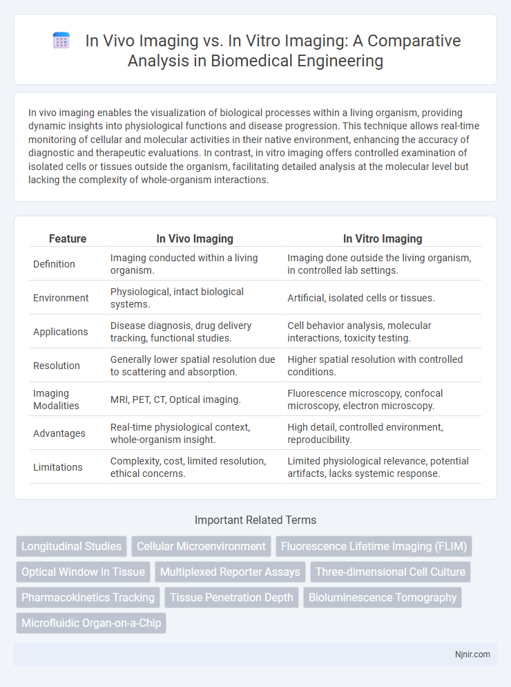
| Feature | In Vivo Imaging | In Vitro Imaging |
|---|---|---|
| Definition | Imaging conducted within a living organism. | Imaging done outside the living organism, in controlled lab settings. |
| Environment | Physiological, intact biological systems. | Artificial, isolated cells or tissues. |
| Applications | Disease diagnosis, drug delivery tracking, functional studies. | Cell behavior analysis, molecular interactions, toxicity testing. |
| Resolution | Generally lower spatial resolution due to scattering and absorption. | Higher spatial resolution with controlled conditions. |
| Imaging Modalities | MRI, PET, CT, Optical imaging. | Fluorescence microscopy, confocal microscopy, electron microscopy. |
| Advantages | Real-time physiological context, whole-organism insight. | High detail, controlled environment, reproducibility. |
| Limitations | Complexity, cost, limited resolution, ethical concerns. | Limited physiological relevance, potential artifacts, lacks systemic response. |
Introduction to Biomedical Imaging Modalities
Biomedical imaging modalities encompass a range of techniques for visualizing biological processes either within living organisms (in vivo) or outside them in controlled environments (in vitro). In vivo imaging provides real-time insights into physiological and pathological conditions by capturing dynamic processes in tissues and organs, utilizing modalities such as MRI, PET, and fluorescence imaging. Conversely, in vitro imaging techniques, including microscopy and flow cytometry, enable detailed cellular and molecular analysis under laboratory conditions, facilitating precise biochemical and genetic investigations.
Defining In Vivo Imaging: Techniques and Applications
In vivo imaging involves non-invasive techniques that visualize biological processes within a living organism, using methods such as MRI, PET, and fluorescence imaging to monitor cellular activities in real-time. This approach enables dynamic study of physiological functions, disease progression, and drug responses in their natural context, providing crucial insights for biomedical research and clinical diagnostics. Applications of in vivo imaging span oncology, neurology, and cardiology, facilitating precise localization of tumors, assessment of brain function, and evaluation of cardiovascular health.
Understanding In Vitro Imaging: Methods and Models
In vitro imaging employs techniques such as fluorescence microscopy, confocal microscopy, and electron microscopy to visualize cellular and molecular processes within controlled laboratory environments. This method utilizes cell cultures, tissue slices, and organoids as models to study biological functions, drug interactions, and disease mechanisms with high spatial resolution. In vitro imaging allows for precise manipulation and real-time monitoring of cellular activities, providing critical insights for biomedical research and pharmaceutical development.
Key Differences Between In Vivo and In Vitro Imaging
In vivo imaging captures biological processes within a living organism, enabling real-time observation of complex interactions in tissues and organs, while in vitro imaging examines isolated cells or tissues under controlled laboratory conditions. In vivo techniques, such as MRI and PET scans, provide dynamic, physiological context but face limitations in resolution and accessibility compared to high-resolution microscopy often used in vitro. Understanding these key differences guides researchers in selecting appropriate imaging methods for studying cellular mechanisms or whole-body biological phenomena.
Resolution and Sensitivity: Imaging Performance Comparison
In vivo imaging offers dynamic, real-time visualization of biological processes within living organisms but often faces challenges with lower resolution and sensitivity due to tissue scattering and absorption. In contrast, in vitro imaging provides higher resolution and sensitivity by utilizing controlled environments and optimized staining or labeling techniques, enabling detailed cellular and molecular analysis. The trade-off between physiological relevance in vivo and enhanced imaging performance in vitro dictates the choice based on specific research objectives.
Biological Relevance: Translational Value in Biomedical Research
In vivo imaging preserves the complex biological environment, providing higher translational value by accurately reflecting physiological and pathological processes within living organisms. In contrast, in vitro imaging offers controlled conditions for detailed cellular and molecular analysis but may lack biological relevance due to absence of systemic interactions. The integration of both methods enhances biomedical research by balancing detailed mechanistic insights with real-time physiological context.
Technological Advances in In Vivo and In Vitro Imaging
Technological advances in in vivo imaging include high-resolution multimodal techniques such as PET-MRI and two-photon microscopy, enabling real-time visualization of biological processes within living organisms. In vitro imaging has progressed with innovations like super-resolution microscopy and high-content screening systems, allowing detailed analysis of cellular structures and molecular interactions in controlled environments. These advancements collectively enhance the precision, resolution, and temporal dynamics of biomedical imaging across both in vivo and in vitro platforms.
Ethical Considerations and Limitations
In vivo imaging involves studying biological processes within living organisms, offering dynamic insights but raising ethical concerns related to animal welfare and the necessity of minimizing harm and distress. In vitro imaging, conducted on cells or tissues outside their natural environment, circumvents many ethical issues but may lack the complexity and physiological relevance of whole-organism studies. Limitations of in vivo imaging include higher costs and regulatory restrictions, whereas in vitro imaging struggles with accurately replicating in vivo conditions and systemic interactions.
Future Trends in Biomedical Imaging
Future trends in biomedical imaging emphasize advancements in in vivo imaging techniques, such as real-time molecular imaging and multi-modal approaches that enhance non-invasive disease monitoring and personalized medicine. In vitro imaging continues to evolve with high-throughput screening and super-resolution microscopy, enabling detailed cellular and molecular analyses critical for drug discovery and biomarker identification. Integration of artificial intelligence and machine learning is expected to revolutionize both in vivo and in vitro imaging by improving image analysis, diagnostic accuracy, and predictive modeling.
Conclusion: Choosing the Optimal Imaging Approach
Selecting the optimal imaging approach depends on the research objective, with in vivo imaging offering dynamic insights into biological processes within living organisms, and in vitro imaging providing controlled environments for detailed molecular and cellular analysis. In vivo imaging techniques, such as MRI and PET, enable real-time visualization of physiological functions, whereas in vitro methods like fluorescence microscopy facilitate high-resolution examination of isolated cells and tissues. Balancing these approaches enhances experimental accuracy, ensuring comprehensive understanding in biomedical studies.
Longitudinal Studies
In vivo imaging enables longitudinal studies by allowing repeated, non-invasive monitoring of biological processes within living organisms, whereas in vitro imaging is limited to static observations of isolated cells or tissues outside their natural environment.
Cellular Microenvironment
In vivo imaging captures dynamic cellular microenvironment interactions within living organisms, providing more physiologically relevant data compared to in vitro imaging, which isolates cells and lacks complex tissue context.
Fluorescence Lifetime Imaging (FLIM)
Fluorescence Lifetime Imaging (FLIM) offers in vivo imaging advantages such as real-time tissue analysis and physiological context, while in vitro FLIM provides precise molecular interactions and controlled environment measurements.
Optical Window in Tissue
In vivo imaging utilizes the optical window in tissue, typically between 650 and 900 nm, to achieve deeper light penetration and clearer visualization of biological structures compared to in vitro imaging, which operates outside living organisms without the constraints of tissue absorption and scattering.
Multiplexed Reporter Assays
Multiplexed reporter assays in vivo imaging enable real-time monitoring of multiple biological processes within living organisms, whereas in vitro imaging provides controlled, high-throughput analysis of reporter signals in isolated cell cultures.
Three-dimensional Cell Culture
Three-dimensional cell culture enables more accurate in vivo imaging by replicating the natural cellular environment, while in vitro imaging typically involves two-dimensional cultures that lack spatial complexity.
Pharmacokinetics Tracking
In vivo imaging enables real-time pharmacokinetics tracking by visualizing drug distribution and metabolism within living organisms, whereas in vitro imaging provides controlled, cellular-level insights but lacks dynamic systemic context.
Tissue Penetration Depth
In vivo imaging achieves greater tissue penetration depth compared to in vitro imaging by utilizing advanced techniques like MRI and PET, enabling real-time visualization of physiological processes within intact organisms.
Bioluminescence Tomography
Bioluminescence tomography enables precise three-dimensional in vivo imaging of molecular and cellular processes, surpassing the spatial limitations of in vitro imaging techniques.
Microfluidic Organ-on-a-Chip
Microfluidic organ-on-a-chip technology enhances in vitro imaging by replicating in vivo physiological conditions, enabling high-resolution, real-time visualization of cellular processes within controlled microenvironments.
In Vivo Imaging vs In Vitro Imaging Infographic