NIR imaging offers deeper tissue penetration and reduced autofluorescence compared to conventional fluorescence imaging, enhancing the clarity of biomedical diagnostics. Fluorescence imaging provides high sensitivity and specificity for molecular targets but is limited by shallow penetration and background noise. Combining NIR imaging with fluorescence techniques advances real-time, non-invasive monitoring of biological processes at cellular and tissue levels.
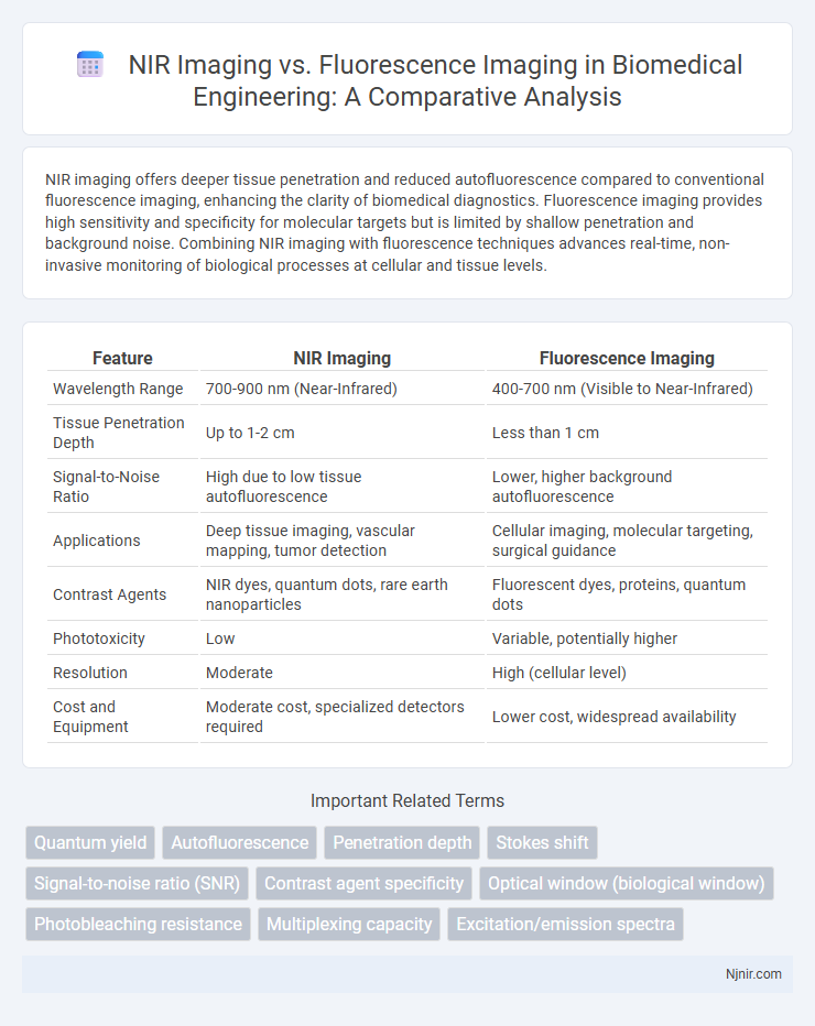
Table of Comparison
| Feature | NIR Imaging | Fluorescence Imaging |
|---|---|---|
| Wavelength Range | 700-900 nm (Near-Infrared) | 400-700 nm (Visible to Near-Infrared) |
| Tissue Penetration Depth | Up to 1-2 cm | Less than 1 cm |
| Signal-to-Noise Ratio | High due to low tissue autofluorescence | Lower, higher background autofluorescence |
| Applications | Deep tissue imaging, vascular mapping, tumor detection | Cellular imaging, molecular targeting, surgical guidance |
| Contrast Agents | NIR dyes, quantum dots, rare earth nanoparticles | Fluorescent dyes, proteins, quantum dots |
| Phototoxicity | Low | Variable, potentially higher |
| Resolution | Moderate | High (cellular level) |
| Cost and Equipment | Moderate cost, specialized detectors required | Lower cost, widespread availability |
Introduction to Biomedical Imaging Techniques
NIR imaging utilizes near-infrared light to penetrate tissues, offering deep tissue visualization with minimal scattering and low background noise. Fluorescence imaging detects emitted light from fluorescent probes activated by specific wavelengths, enabling high specificity and contrast for targeting molecular markers. Both techniques are pivotal in biomedical imaging, with NIR providing enhanced depth and fluorescence imaging delivering precise molecular-level information.
Principles of NIR Imaging
NIR imaging operates on the principle of using near-infrared light, typically between 700 and 900 nm wavelengths, to penetrate biological tissues with minimal absorption and scattering, enabling deeper tissue visualization compared to visible light. This imaging technique relies on the reflectance, absorption, or emission of NIR light by tissues or contrast agents, allowing for high-contrast images of vascular structures, oxygenation levels, and molecular targets. The intrinsic low autofluorescence and reduced photodamage in the NIR window enhance the sensitivity and specificity of NIR imaging for in vivo diagnostics.
Fundamentals of Fluorescence Imaging
Fluorescence imaging relies on the excitation of fluorophores using specific wavelengths of light, leading to the emission of light at longer wavelengths, which is then detected to create an image. This technique provides high sensitivity and specificity for molecular and cellular visualization by exploiting the unique emission spectra of fluorescent markers. In contrast, Near-Infrared (NIR) imaging primarily captures reflected or emitted light in the NIR spectrum, offering deeper tissue penetration but generally lower specificity than fluorescence imaging.
Comparative Mechanisms: NIR vs Fluorescence
NIR imaging relies on near-infrared light absorption and reflection to capture deeper tissue structures due to its longer wavelengths, whereas fluorescence imaging detects emitted light from fluorescent probes excited by specific wavelengths, allowing for high specificity in molecular targeting. NIR imaging offers better penetration depth and reduced autofluorescence interference compared to fluorescence imaging, which is constrained by limited tissue penetration and background noise. The distinct mechanisms of NIR absorption versus fluorescence emission enable complementary applications in biomedical imaging, with NIR excelling in structural visualization and fluorescence providing molecular-level insights.
Applications in Disease Diagnosis
NIR imaging offers deep tissue penetration and low background autofluorescence, making it ideal for detecting tumors and vascular abnormalities in disease diagnosis. Fluorescence imaging provides high sensitivity and specificity through targeted molecular probes, enabling precise visualization of cancer cells and infectious agents. Combining both methods enhances diagnostic accuracy by leveraging NIR's depth with fluorescence's molecular specificity in real-time clinical applications.
Imaging Depth and Resolution Considerations
Near-Infrared (NIR) imaging offers greater tissue penetration with depths up to several centimeters, enabling visualization of structures beyond surface layers, while fluorescence imaging typically achieves high spatial resolution but at shallower depths due to light scattering and absorption. NIR imaging benefits from reduced scattering and autofluorescence, enhancing imaging depth but sometimes at the expense of resolution compared to fluorescence techniques which provide finer detail for superficial tissues. Choosing between these modalities depends on the required balance between imaging depth and resolution for specific biomedical applications.
Sensitivity and Specificity Analysis
Near-Infrared (NIR) imaging offers higher tissue penetration and reduced scattering, resulting in enhanced sensitivity for detecting deep or weak signals compared to fluorescence imaging. Fluorescence imaging, while highly sensitive to specific molecular targets due to its direct emission detection, often suffers from lower specificity caused by autofluorescence background and overlapping spectra. Sensitivity and specificity metrics favor NIR imaging in applications requiring deep tissue visualization, whereas fluorescence imaging excels in targeted molecular specificity within shallow or surface tissues.
Clinical Advantages and Limitations
Near-infrared (NIR) imaging offers deep tissue penetration and low autofluorescence, enabling high-contrast visualization of vascular structures and tumors in clinical settings. Fluorescence imaging provides real-time molecular and cellular-level detail, enhancing surgical precision and tumor margin detection but is limited by shallower tissue penetration and potential photobleaching. Both modalities are complementary, with NIR suited for broader structural imaging and fluorescence excelling in cellular specificity, although fluorescence requires careful management of dye toxicity and imaging duration.
Recent Advances and Future Trends
Recent advances in NIR imaging have focused on improving tissue penetration depth and reducing background noise through the development of new contrast agents and enhanced detector sensitivity. Fluorescence imaging has seen significant progress with the introduction of near-infrared fluorophores and multimodal probes, enabling higher resolution and real-time molecular tracking in clinical diagnostics. Future trends indicate a convergence of NIR and fluorescence techniques with AI-driven image analysis and wearable imaging devices to facilitate personalized medicine and non-invasive monitoring.
Conclusion: Choosing the Optimal Imaging Modality
NIR imaging provides deeper tissue penetration and lower background autofluorescence, making it ideal for in vivo applications requiring high spatial resolution. Fluorescence imaging offers higher sensitivity and multiplexing capabilities, suitable for detailed cellular and molecular analysis. Selecting the optimal modality depends on balancing depth, resolution, sensitivity, and the specific biological context of the study.
Quantum yield
NIR imaging offers deeper tissue penetration but generally exhibits lower quantum yield compared to fluorescence imaging, which provides higher quantum yield for brighter signal intensity in biological applications.
Autofluorescence
NIR imaging reduces interference from tissue autofluorescence, providing higher contrast and deeper tissue penetration compared to fluorescence imaging, which is often limited by strong background signals from endogenous fluorophores.
Penetration depth
NIR imaging offers greater tissue penetration depth up to several centimeters compared to fluorescence imaging, which is generally limited to a few millimeters due to light scattering and absorption.
Stokes shift
Near-Infrared (NIR) imaging benefits from a larger Stokes shift compared to fluorescence imaging, resulting in reduced background noise and enhanced signal clarity for deeper tissue visualization.
Signal-to-noise ratio (SNR)
NIR imaging offers a higher signal-to-noise ratio (SNR) compared to fluorescence imaging due to reduced tissue autofluorescence and deeper light penetration, enhancing image clarity and detection sensitivity.
Contrast agent specificity
NIR imaging utilizes contrast agents with deeper tissue penetration and lower autofluorescence, while fluorescence imaging relies on highly specific contrast agents targeting particular molecular markers for enhanced signal specificity.
Optical window (biological window)
NIR imaging exploits the biological optical window (650-950 nm) to achieve deeper tissue penetration and reduced autofluorescence compared to fluorescence imaging predominantly operating outside this range.
Photobleaching resistance
NIR imaging exhibits superior photobleaching resistance compared to fluorescence imaging, enabling longer-lasting signal stability for extended biomedical visualization.
Multiplexing capacity
Near-infrared (NIR) imaging offers limited multiplexing capacity due to spectral overlap, while fluorescence imaging enables higher multiplexing through distinct fluorophore emission spectra and advanced filter technologies.
Excitation/emission spectra
NIR imaging uses near-infrared light typically between 700-900 nm for excitation and emission, enabling deeper tissue penetration and reduced autofluorescence compared to fluorescence imaging, which employs a broader excitation spectrum (400-700 nm) and visible emission, resulting in higher background noise and limited tissue depth.
NIR imaging vs Fluorescence imaging Infographic