AI imaging analysis in biomedical engineering enhances diagnostic accuracy by rapidly processing complex medical images, identifying subtle patterns that may be overlooked in manual radiology reads. It reduces human error and enables quantifiable, reproducible results, facilitating early disease detection and personalized treatment planning. While manual radiology relies on expert interpretation, AI integration streamlines workflows, increases throughput, and supports more consistent decision-making in clinical settings.
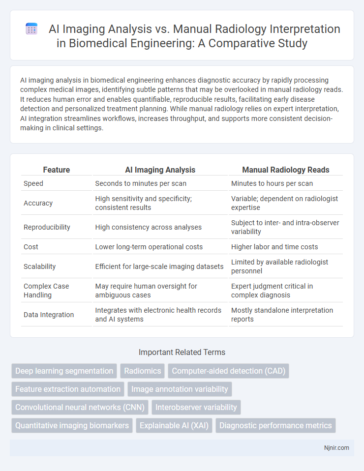
Table of Comparison
| Feature | AI Imaging Analysis | Manual Radiology Reads |
|---|---|---|
| Speed | Seconds to minutes per scan | Minutes to hours per scan |
| Accuracy | High sensitivity and specificity; consistent results | Variable; dependent on radiologist expertise |
| Reproducibility | High consistency across analyses | Subject to inter- and intra-observer variability |
| Cost | Lower long-term operational costs | Higher labor and time costs |
| Scalability | Efficient for large-scale imaging datasets | Limited by available radiologist personnel |
| Complex Case Handling | May require human oversight for ambiguous cases | Expert judgment critical in complex diagnosis |
| Data Integration | Integrates with electronic health records and AI systems | Mostly standalone interpretation reports |
Introduction to AI Imaging Analysis in Biomedical Engineering
AI imaging analysis in biomedical engineering leverages advanced algorithms and deep learning models to enhance the accuracy and efficiency of medical image interpretation compared to manual radiology reads. This technology enables rapid identification of abnormalities and quantification of disease markers in modalities such as MRI, CT, and X-ray scans, reducing human error and inter-observer variability. Integration of AI tools supports radiologists by providing data-driven insights and automated measurements, accelerating diagnosis and improving patient outcomes.
Evolution of Manual Radiology Reads
Manual radiology reads have evolved significantly with advancements in imaging technology and increased specialization, enhancing diagnostic accuracy and efficiency. Radiologists now integrate complex imaging modalities like MRI, CT, and PET scans, applying refined protocols and standardized reporting systems such as BI-RADS and LI-RADS. Continuous education and AI-assisted tools have further improved interpretative skills, yet manual reads rely on radiologist expertise for nuances and clinical context that AI imaging analysis continues to complement.
Key Differences Between AI Imaging and Manual Interpretation
AI imaging analysis utilizes advanced algorithms and deep learning models to quickly process large volumes of medical images, enhancing detection accuracy and consistency compared to manual radiology reads. Manual interpretation relies heavily on radiologists' expertise and experience, which can introduce variability and longer turnaround times due to human limitations. Key differences include AI's ability to standardize assessments, improve diagnostic speed, and detect subtle patterns that might be missed in manual reads.
Accuracy and Diagnostic Performance Comparison
AI imaging analysis demonstrates higher accuracy and consistency in detecting abnormalities compared to manual radiology reads, reducing human error and inter-observer variability. Studies reveal AI algorithms achieve sensitivity and specificity rates exceeding 90% in diagnosing conditions such as lung nodules and breast cancer, often outperforming traditional methods. Enhanced diagnostic performance with AI supports faster decision-making and improved patient outcomes by enabling early and precise detection.
Workflow Efficiency: AI vs. Human Radiologists
AI imaging analysis dramatically enhances workflow efficiency by rapidly processing vast amounts of data with consistent accuracy, reducing the time needed for image interpretation. Human radiologists, while skilled in nuanced decision-making and complex cases, often face bottlenecks due to workload and variability in reading times. Integrating AI assists radiologists by prioritizing cases and flagging abnormalities, streamlining their workflow and improving overall diagnostic throughput.
Integration Challenges and Adaptation in Clinical Settings
AI imaging analysis offers rapid, consistent evaluations but faces integration challenges such as interoperability with existing PACS systems and resistance from radiologists accustomed to manual reads. Manual radiology expertise provides nuanced interpretation and clinical judgment but struggles with time constraints and variability in accuracy. Successful adaptation in clinical settings requires seamless workflow integration, user training, and validation studies to build trust and optimize diagnostic efficiency.
Cost Implications and Resource Allocation
AI imaging analysis significantly reduces cost implications by automating the interpretation of radiological images, thereby decreasing the need for extensive human labor and minimizing errors that lead to costly diagnostic delays. Manual radiology reads require substantial investment in skilled radiologists and prolonged turnaround times, which strain healthcare resources and increase operational expenses. Integrating AI systems streamlines resource allocation, enabling radiologists to focus on complex cases while optimizing workflow efficiency and reducing overall imaging costs.
Ethical Considerations in Automated Imaging Analysis
AI imaging analysis in radiology raises ethical considerations including data privacy, informed consent, and potential algorithmic bias affecting diagnostic accuracy. Ensuring transparency in AI decision-making processes and maintaining patient trust requires rigorous validation and continuous monitoring of automated systems. Balancing efficiency gains with accountability safeguards is critical to ethical integration of AI in clinical imaging workflows.
Case Studies: AI Successes and Limitations
Case studies reveal AI imaging analysis excels in detecting subtle abnormalities with speed and consistency, significantly reducing diagnostic errors in large-scale screenings. However, limitations persist in complex cases requiring nuanced interpretation, where manual radiology reads demonstrate superior contextual judgment and adaptability. Combining AI capabilities with expert radiologists enhances diagnostic accuracy and streamlines workflows, optimizing patient outcomes.
Future Directions in Biomedical Imaging Technologies
AI imaging analysis outperforms manual radiology reads in speed and accuracy, leveraging deep learning algorithms to detect subtle patterns beyond human perception. Future directions in biomedical imaging emphasize integrating AI with multimodal data, enhancing diagnostic precision and personalized treatment planning. Advancements in real-time image processing and explainable AI models will drive more reliable, interpretable clinical decision support systems.
Deep learning segmentation
Deep learning segmentation in AI imaging analysis significantly improves accuracy and efficiency compared to manual radiology reads by automating the detection and quantification of complex anatomical structures.
Radiomics
Radiomics enhances AI imaging analysis by extracting quantitative features from medical images, offering higher reproducibility and predictive accuracy compared to manual radiology reads.
Computer-aided detection (CAD)
Computer-aided detection (CAD) systems significantly enhance AI imaging analysis by improving accuracy and efficiency in identifying abnormalities compared to manual radiology reads.
Feature extraction automation
AI imaging analysis automates feature extraction with higher accuracy and efficiency compared to manual radiology reads, significantly reducing diagnostic time and human error.
Image annotation variability
AI imaging analysis reduces image annotation variability by providing consistent and quantifiable assessments compared to the subjective interpretations and inter-reader variability seen in manual radiology reads.
Convolutional neural networks (CNN)
Convolutional neural networks (CNN) significantly enhance AI imaging analysis by providing faster, more accurate detection and diagnosis compared to traditional manual radiology reads, reducing human error and increasing diagnostic consistency.
Interobserver variability
AI imaging analysis reduces interobserver variability significantly compared to manual radiology reads by providing consistent and standardized interpretations across diverse cases.
Quantitative imaging biomarkers
AI imaging analysis enhances the accuracy and efficiency of quantitative imaging biomarker extraction compared to manual radiology reads by enabling standardized, reproducible, and high-throughput assessments.
Explainable AI (XAI)
Explainable AI (XAI) in imaging analysis enhances diagnostic transparency and accuracy by providing interpretable insights that complement manual radiology reads, improving clinical decision-making and trust.
Diagnostic performance metrics
AI imaging analysis demonstrates higher sensitivity and specificity in diagnostic performance metrics compared to manual radiology reads, significantly improving accuracy and reducing false positives and negatives.
AI imaging analysis vs Manual radiology reads Infographic