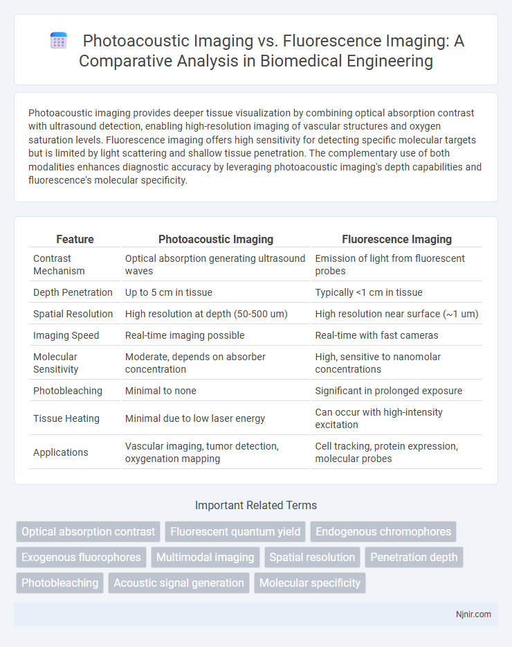
Photoacoustic imaging provides deeper tissue visualization by combining optical absorption contrast with ultrasound detection, enabling high-resolution imaging of vascular structures and oxygen saturation levels. Fluorescence imaging offers high sensitivity for detecting specific molecular targets but is limited by light scattering and shallow tissue penetration. The complementary use of both modalities enhances diagnostic accuracy by leveraging photoacoustic imaging's depth capabilities and fluorescence's molecular specificity.
Table of Comparison
| Feature | Photoacoustic Imaging | Fluorescence Imaging |
|---|---|---|
| Contrast Mechanism | Optical absorption generating ultrasound waves | Emission of light from fluorescent probes |
| Depth Penetration | Up to 5 cm in tissue | Typically <1 cm in tissue |
| Spatial Resolution | High resolution at depth (50-500 um) | High resolution near surface (~1 um) |
| Imaging Speed | Real-time imaging possible | Real-time with fast cameras |
| Molecular Sensitivity | Moderate, depends on absorber concentration | High, sensitive to nanomolar concentrations |
| Photobleaching | Minimal to none | Significant in prolonged exposure |
| Tissue Heating | Minimal due to low laser energy | Can occur with high-intensity excitation |
| Applications | Vascular imaging, tumor detection, oxygenation mapping | Cell tracking, protein expression, molecular probes |
Introduction to Biomedical Imaging Modalities
Photoacoustic imaging combines optical absorption contrast with ultrasonic resolution, enabling deeper tissue visualization compared to fluorescence imaging, which relies on emitted light from fluorophores and is limited by scattering and absorption in biological tissues. Biomedical imaging modalities like photoacoustic imaging offer enhanced spatial resolution and functional information by detecting ultrasonic waves generated from absorbed light, whereas fluorescence imaging provides high sensitivity for molecular and cellular level detection but with limited tissue penetration. These complementary techniques address different challenges in biomedical imaging, optimizing diagnostic capabilities across various clinical and research applications.
Principles of Photoacoustic Imaging
Photoacoustic imaging operates by converting absorbed pulsed laser light into ultrasonic waves through thermoelastic expansion, enabling high-resolution visualization of optical absorption contrast in tissues. This technique detects ultrasound signals generated by transient temperature rises in endogenous chromophores like hemoglobin, providing deep tissue imaging beyond the optical diffusion limit. Unlike fluorescence imaging that relies on emitted light from excited fluorophores, photoacoustic imaging combines optical contrast with ultrasonic spatial resolution, making it effective for functional and molecular imaging in biomedical applications.
Fundamentals of Fluorescence Imaging
Fluorescence imaging relies on the excitation of fluorophores by specific wavelengths of light, causing them to emit light at a longer wavelength for detection. This technique offers high sensitivity and spatial resolution by capturing emitted photons from labeled biological structures or molecules. Compared to photoacoustic imaging, fluorescence imaging provides direct visualization of molecular events but is limited by shallow tissue penetration due to light scattering and absorption.
Comparative Sensitivity and Specificity
Photoacoustic imaging offers higher sensitivity by detecting ultrasonic waves generated from optical absorption, enabling deeper tissue visualization compared to fluorescence imaging, which relies on emitted light with limited penetration. Specificity in photoacoustic imaging often surpasses fluorescence methods due to its ability to distinguish between different chromophores based on their unique absorption spectra. Fluorescence imaging provides high molecular specificity through targeted fluorophores but experiences lower sensitivity in deep tissues due to light scattering and absorption.
Depth Penetration Capabilities
Photoacoustic imaging offers superior depth penetration capabilities compared to fluorescence imaging, typically reaching several centimeters in biological tissues due to its use of ultrasonic waves generated by light absorption. Fluorescence imaging is limited to superficial depths of a few millimeters because of light scattering and absorption in tissues. This makes photoacoustic imaging more suitable for deep-tissue visualization in medical diagnostics and research.
Spatial and Temporal Resolution Differences
Photoacoustic imaging offers higher spatial resolution at greater tissue depths, typically achieving resolutions of 50-200 micrometers compared to fluorescence imaging's 5-20 micrometers limited to superficial layers. Temporal resolution in photoacoustic imaging can reach real-time video rates (up to 50 Hz) due to rapid acoustic wave detection, whereas fluorescence imaging generally exhibits slower acquisition speeds constrained by slower photon emission and camera exposure times. These differences make photoacoustic imaging more suitable for dynamic deep-tissue visualization, while fluorescence imaging excels in high-resolution surface-level observations.
Contrast Agents: Types and Biocompatibility
Photoacoustic imaging utilizes contrast agents such as gold nanoparticles, carbon nanotubes, and organic dyes that exhibit strong optical absorption to generate high-resolution, deep-tissue images, with a focus on biocompatibility and minimal toxicity for clinical applications. Fluorescence imaging employs contrast agents like organic fluorophores, quantum dots, and fluorescent proteins, optimized for brightness and wavelength specificity but often limited by photobleaching and potential cytotoxicity. Both modalities prioritize biocompatible formulations, yet photoacoustic agents generally offer greater stability and deeper tissue penetration compared to fluorescence agents, making them suitable for in vivo imaging with enhanced safety profiles.
Clinical Applications and Case Studies
Photoacoustic imaging provides high-resolution, deep-tissue visualization by detecting ultrasonic waves generated from light absorption, making it valuable in oncology for tumor margin delineation and vascular imaging. Fluorescence imaging excels in real-time intraoperative guidance through targeted fluorescent probes, widely used in sentinel lymph node mapping and cancer detection. Case studies highlight photoacoustic imaging's success in identifying breast cancer vasculature while fluorescence imaging demonstrates efficacy in guiding tumor resection in brain surgeries.
Limitations and Technical Challenges
Photoacoustic imaging faces limitations related to depth penetration and spatial resolution due to optical scattering and acoustic attenuation in biological tissues. Fluorescence imaging struggles with photobleaching, autofluorescence background noise, and limited depth penetration caused by light absorption and scattering. Both modalities encounter challenges in achieving high contrast and specificity, with photoacoustic imaging requiring complex signal processing and fluorescence imaging dependent on stable and biocompatible fluorophores.
Future Directions in Hybrid Imaging Technologies
Photoacoustic imaging offers deep tissue penetration and high spatial resolution, complementing fluorescence imaging's sensitivity to molecular markers. Future directions in hybrid imaging technologies emphasize integrating these modalities to enhance diagnostic accuracy and real-time monitoring in oncology and vascular diseases. Advances in multimodal contrast agents and machine learning algorithms will drive the development of compact, cost-effective hybrid imaging systems for clinical and preclinical applications.
Optical absorption contrast
Photoacoustic imaging leverages strong optical absorption contrast of endogenous chromophores like hemoglobin, enabling deeper tissue visualization than fluorescence imaging, which relies on weaker fluorescence emission with limited penetration depth.
Fluorescent quantum yield
Fluorescence imaging relies heavily on fluorescent quantum yield, a key parameter measuring the efficiency of photon emission that directly influences the brightness and sensitivity of imaging compared to photoacoustic imaging.
Endogenous chromophores
Photoacoustic imaging provides higher contrast and deeper tissue penetration by detecting endogenous chromophores such as hemoglobin and melanin, whereas fluorescence imaging primarily relies on exogenous fluorophores with limited depth sensitivity.
Exogenous fluorophores
Photoacoustic imaging offers deeper tissue penetration and higher spatial resolution compared to fluorescence imaging, especially when utilizing exogenous fluorophores with strong optical absorption properties for enhanced signal contrast.
Multimodal imaging
Photoacoustic imaging offers high-resolution, deep-tissue visualization by detecting ultrasound signals generated from optical absorption, while fluorescence imaging provides high sensitivity in molecular detection, making their combination in multimodal imaging highly effective for comprehensive diagnostic accuracy and functional-anatomical correlation.
Spatial resolution
Photoacoustic imaging offers higher spatial resolution in deep tissue compared to fluorescence imaging, which is limited by light scattering and absorption.
Penetration depth
Photoacoustic imaging achieves greater penetration depth in biological tissues, reaching several centimeters, compared to fluorescence imaging, which is typically limited to a few millimeters.
Photobleaching
Photoacoustic imaging offers superior resistance to photobleaching compared to fluorescence imaging, enabling longer imaging sessions with enhanced signal stability.
Acoustic signal generation
Photoacoustic imaging generates acoustic signals through thermoelastic expansion induced by pulsed laser absorption, enabling deeper tissue penetration and high-resolution imaging compared to the photon emission-based signal detection in fluorescence imaging.
Molecular specificity
Photoacoustic imaging offers higher molecular specificity by detecting optical absorption contrasts of endogenous chromophores, whereas fluorescence imaging relies on external fluorescent probes with limited tissue penetration and potential background autofluorescence.
Photoacoustic imaging vs Fluorescence imaging Infographic