Functional MRI (fMRI) measures brain activity by detecting changes in blood oxygenation, providing insights into neural function and brain mapping. Diffusion MRI (dMRI) captures the diffusion of water molecules along white matter tracts, enabling detailed visualization of neural pathways and structural connectivity. These complementary imaging techniques enhance understanding of brain function and architecture in biomedical engineering applications.
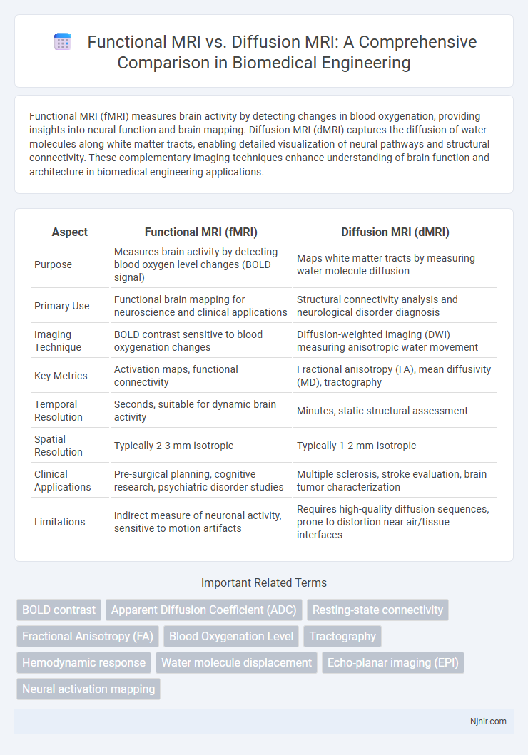
Table of Comparison
| Aspect | Functional MRI (fMRI) | Diffusion MRI (dMRI) |
|---|---|---|
| Purpose | Measures brain activity by detecting blood oxygen level changes (BOLD signal) | Maps white matter tracts by measuring water molecule diffusion |
| Primary Use | Functional brain mapping for neuroscience and clinical applications | Structural connectivity analysis and neurological disorder diagnosis |
| Imaging Technique | BOLD contrast sensitive to blood oxygenation changes | Diffusion-weighted imaging (DWI) measuring anisotropic water movement |
| Key Metrics | Activation maps, functional connectivity | Fractional anisotropy (FA), mean diffusivity (MD), tractography |
| Temporal Resolution | Seconds, suitable for dynamic brain activity | Minutes, static structural assessment |
| Spatial Resolution | Typically 2-3 mm isotropic | Typically 1-2 mm isotropic |
| Clinical Applications | Pre-surgical planning, cognitive research, psychiatric disorder studies | Multiple sclerosis, stroke evaluation, brain tumor characterization |
| Limitations | Indirect measure of neuronal activity, sensitive to motion artifacts | Requires high-quality diffusion sequences, prone to distortion near air/tissue interfaces |
Introduction to Functional MRI and Diffusion MRI
Functional MRI (fMRI) measures brain activity by detecting changes in blood oxygenation levels, providing insights into neural function and brain regions involved in specific tasks. Diffusion MRI, including Diffusion Tensor Imaging (DTI), maps the diffusion of water molecules in tissue, revealing the microstructural integrity and connectivity of white matter pathways. These techniques complement each other by combining functional brain mapping with detailed structural connectivity analysis for comprehensive neuroimaging.
Principles of Functional MRI (fMRI)
Functional MRI (fMRI) operates by detecting changes in blood oxygenation levels through the Blood Oxygen Level Dependent (BOLD) signal, which reflects neural activity in specific brain regions. This technique measures hemodynamic responses triggered by neuronal activation, providing high spatial resolution maps of brain function. Unlike Diffusion MRI, which tracks the movement of water molecules to visualize neural pathways, fMRI emphasizes functional brain activity patterns.
Principles of Diffusion MRI (dMRI)
Diffusion MRI (dMRI) exploits the random Brownian motion of water molecules to map tissue microstructure, particularly in neural pathways, by measuring the diffusion of water in different directions. Unlike functional MRI (fMRI), which detects brain activity through blood oxygen level-dependent (BOLD) signals, dMRI provides detailed visualization of white matter tracts using techniques such as diffusion tensor imaging (DTI) to quantify anisotropic diffusion. This enables the assessment of neural connectivity and microstructural integrity critical for understanding brain organization and diagnosing various neurological disorders.
Imaging Techniques and Data Acquisition
Functional MRI (fMRI) measures brain activity by detecting changes in blood oxygenation and flow, utilizing blood-oxygen-level-dependent (BOLD) contrast to capture dynamic neural responses with high temporal resolution. Diffusion MRI (dMRI) maps white matter pathways by tracking the diffusion of water molecules in tissue, employing diffusion-weighted imaging (DWI) sequences that provide detailed microstructural information with high spatial resolution. While fMRI data acquisition focuses on repeated volumetric scans to monitor brain function over time, dMRI requires multiple gradient directions to reconstruct diffusion tensors, enabling mapping of neural connectivity and fiber orientation.
Biological Basis of Signal Generation
Functional MRI (fMRI) detects brain activity by measuring changes in blood oxygenation levels, specifically the Blood Oxygen Level Dependent (BOLD) signal, which reflects neuronal activation through hemodynamic responses. Diffusion MRI (dMRI) maps the diffusion of water molecules within tissue, capturing microstructural characteristics of neural fibers by measuring the directional movement of water along white matter tracts. The biological basis of fMRI relies on vascular response to neural activity, while dMRI depends on the anisotropic diffusion of water constrained by cellular membranes and fiber architecture.
Key Applications in Biomedical Engineering
Functional MRI (fMRI) maps brain activity by detecting blood flow changes, crucial for neuroimaging studies, brain-computer interfaces, and pre-surgical planning. Diffusion MRI (dMRI) tracks water molecule movement along white matter tracts, enabling detailed analysis of brain connectivity, neural pathway integrity, and early diagnosis of neurodegenerative diseases. Both modalities play pivotal roles in biomedical engineering by advancing diagnostic accuracy and guiding therapeutic interventions in neurological disorders.
Comparative Analysis: Strengths and Limitations
Functional MRI (fMRI) excels at measuring brain activity by detecting changes in blood oxygenation, offering high spatial resolution for mapping functional areas, yet it is limited by its indirect measurement of neural activity and susceptibility to motion artifacts. Diffusion MRI (dMRI) provides detailed insights into white matter microstructure by tracking the diffusion of water molecules, allowing for the visualization of neural pathways but suffers from lower spatial resolution and challenges in resolving complex fiber crossings. While fMRI is optimal for studying dynamic brain function, dMRI is indispensable for structural connectivity analysis, making their combined use crucial for comprehensive neuroimaging.
Clinical and Research Utility
Functional MRI (fMRI) measures brain activity by detecting changes in blood oxygenation levels, making it invaluable for mapping functional areas in clinical settings such as pre-surgical planning and neurological disorder assessment. Diffusion MRI, particularly Diffusion Tensor Imaging (DTI), visualizes white matter tracts by tracking water molecule diffusion, proving essential for diagnosing conditions like stroke, multiple sclerosis, and brain tumors as well as investigating neural connectivity in research. Both modalities complement each other by providing comprehensive insights into brain function and structure, enhancing clinical decision-making and advancing neuroscientific studies.
Emerging Trends and Future Directions
Functional MRI (fMRI) advancements emphasize high-resolution brain activity mapping through enhanced BOLD signal sensitivity and integration with machine learning algorithms for improved cognitive state decoding. Diffusion MRI is evolving with ultra-high gradient systems enabling detailed microstructural imaging, alongside sophisticated tractography methods for precise neural pathway reconstruction. Emerging trends highlight multimodal fusion of fMRI and diffusion MRI to correlate functional connectivity with structural networks, driving personalized neurodiagnostics and targeted therapeutic interventions.
Conclusion: Choosing the Right Modality
Functional MRI (fMRI) excels in mapping brain activity by detecting blood oxygenation changes, crucial for cognitive and neurological studies. Diffusion MRI specializes in visualizing white matter tract integrity and neural connectivity through water molecule diffusion patterns, making it essential for assessing structural brain abnormalities. Selecting the appropriate modality depends on whether the clinical or research focus targets brain function dynamics or structural connectivity.
BOLD contrast
Functional MRI uses BOLD contrast to measure brain activity by detecting changes in blood oxygenation, while Diffusion MRI maps the diffusion of water molecules to visualize neural tissue microstructure.
Apparent Diffusion Coefficient (ADC)
Apparent Diffusion Coefficient (ADC) values derived from Diffusion MRI quantify water molecule movement in tissues, providing distinct microstructural insights compared to Functional MRI's measurement of brain activity through blood oxygen level-dependent signals.
Resting-state connectivity
Resting-state functional MRI (fMRI) measures spontaneous brain activity and functional connectivity by detecting blood oxygen level-dependent (BOLD) signals, whereas diffusion MRI maps structural connectivity by tracking the diffusion of water molecules along white matter tracts.
Fractional Anisotropy (FA)
Fractional Anisotropy (FA) in Diffusion MRI quantifies the directional coherence of water diffusion in white matter, providing insights into neural microstructure that complements the brain activity mapping achieved by Functional MRI.
Blood Oxygenation Level
Functional MRI measures brain activity by detecting changes in blood oxygenation levels, whereas Diffusion MRI maps the diffusion of water molecules without directly reflecting blood oxygenation.
Tractography
Diffusion MRI enables detailed tractography by mapping white matter fiber pathways in the brain, whereas functional MRI measures brain activity through blood oxygen level changes without directly visualizing neural connections.
Hemodynamic response
Functional MRI measures brain activity by detecting changes in the hemodynamic response associated with neural activation, while Diffusion MRI maps the diffusion of water molecules to reveal white matter microstructure without directly reflecting hemodynamic changes.
Water molecule displacement
Functional MRI detects brain activity by measuring blood oxygenation changes, while Diffusion MRI maps the displacement of water molecules to reveal tissue microstructure and connectivity.
Echo-planar imaging (EPI)
Echo-planar imaging (EPI) enables rapid acquisition in both functional MRI (fMRI) to capture blood oxygen level-dependent (BOLD) signals and diffusion MRI to map water molecule movement, making it critical for high-resolution brain activity and microstructural analysis.
Neural activation mapping
Functional MRI precisely maps neural activation by measuring blood oxygen level-dependent changes, while Diffusion MRI visualizes neural connectivity by tracking water molecule movement along white matter tracts.
Functional MRI vs Diffusion MRI Infographic