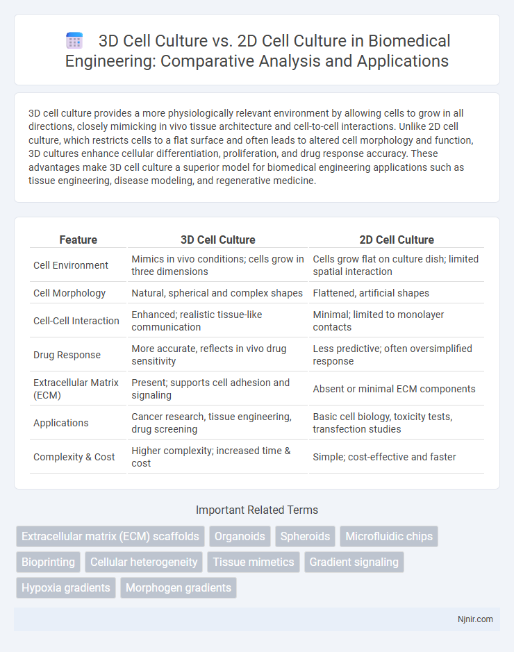
3D cell culture provides a more physiologically relevant environment by allowing cells to grow in all directions, closely mimicking in vivo tissue architecture and cell-to-cell interactions. Unlike 2D cell culture, which restricts cells to a flat surface and often leads to altered cell morphology and function, 3D cultures enhance cellular differentiation, proliferation, and drug response accuracy. These advantages make 3D cell culture a superior model for biomedical engineering applications such as tissue engineering, disease modeling, and regenerative medicine.
Table of Comparison
| Feature | 3D Cell Culture | 2D Cell Culture |
|---|---|---|
| Cell Environment | Mimics in vivo conditions; cells grow in three dimensions | Cells grow flat on culture dish; limited spatial interaction |
| Cell Morphology | Natural, spherical and complex shapes | Flattened, artificial shapes |
| Cell-Cell Interaction | Enhanced; realistic tissue-like communication | Minimal; limited to monolayer contacts |
| Drug Response | More accurate, reflects in vivo drug sensitivity | Less predictive; often oversimplified response |
| Extracellular Matrix (ECM) | Present; supports cell adhesion and signaling | Absent or minimal ECM components |
| Applications | Cancer research, tissue engineering, drug screening | Basic cell biology, toxicity tests, transfection studies |
| Complexity & Cost | Higher complexity; increased time & cost | Simple; cost-effective and faster |
Introduction to 3D and 2D Cell Culture Systems
2D cell culture systems involve growing cells in a flat monolayer on plastic or glass surfaces, offering ease of use and high reproducibility for studying cellular behavior in a simplified environment. In contrast, 3D cell culture systems provide cells with a spatial architecture that closely mimics in vivo tissue, enabling more physiologically relevant interactions and nutrient gradients. Advances in 3D culture techniques, such as spheroids, organoids, and scaffold-based models, enhance cell differentiation, proliferation, and drug response, making them superior for accurate disease modeling and drug screening.
Historical Evolution of Cell Culture Techniques
The historical evolution of cell culture techniques began with 2D cell culture, where cells were grown on flat plastic or glass surfaces, providing a simplified environment to study cellular behavior. The emergence of 3D cell culture revolutionized the field by enabling cells to grow in three-dimensional matrices, better mimicking the in vivo environment and improving physiological relevance in drug testing and disease modeling. Advances in biomaterials and scaffold technology have driven the transition from 2D monolayers to 3D spheroids, organoids, and microtissues, expanding the possibilities for regenerative medicine and cancer research.
Structural Differences Between 2D and 3D Cell Models
3D cell cultures provide a more physiologically relevant environment by enabling cells to grow in all directions, closely mimicking in vivo tissue architecture, unlike 2D cell cultures that restrict cells to a flat, two-dimensional surface. The extracellular matrix in 3D models supports complex cell-cell and cell-matrix interactions, promoting natural cell morphology, differentiation, and signaling pathways, whereas 2D cultures often lead to altered cell shape and limited biological functions. These structural differences significantly impact cellular behavior, drug response, and gene expression, making 3D cultures superior for translational research and drug discovery.
Microenvironment and Cellular Interactions
3D cell culture better mimics the in vivo microenvironment by enabling cells to grow in all directions, facilitating complex cell-cell and cell-matrix interactions that are limited in 2D cultures. This spatial organization in 3D systems promotes more physiologically relevant signaling pathways, gene expression, and cellular behavior. In contrast, 2D cultures restrict cells to a flat surface, often leading to altered morphology and limited extracellular matrix deposition, which can impact experimental outcomes.
Physiological Relevance in Biomedical Research
3D cell culture systems closely mimic the in vivo microenvironment by allowing cells to grow in all directions, promoting natural cell-cell and cell-matrix interactions crucial for accurate physiological relevance in biomedical research. Unlike 2D cultures, which restrict cells to flat surfaces and alter morphologies and functions, 3D cultures provide more predictive models for drug responses, cancer progression, and tissue engineering studies. The enhanced physiological relevance of 3D cultures leads to improved data reliability and translational potential in disease modeling and therapeutic development.
Applications in Drug Screening and Toxicology
3D cell culture models more accurately mimic the physiological environment, enhancing predictive accuracy in drug screening by reflecting cell-cell and cell-matrix interactions absent in 2D cultures. These models improve toxicity assessments by exhibiting more relevant metabolic and diffusion gradients, leading to better identification of drug-induced cytotoxicity and side effects. Consequently, 3D cultures are increasingly preferred for high-throughput screening and toxicological studies in pharmaceutical development.
Advantages and Limitations of 2D Cell Cultures
2D cell cultures offer simplicity, cost-effectiveness, and ease of observation, making them suitable for high-throughput screening and basic cellular studies. However, their limitations include lack of realistic cell-cell and cell-matrix interactions, leading to altered cell morphology and function compared to in vivo conditions. These constraints often result in reduced physiological relevance and predictive accuracy for drug development and disease modeling.
Advantages and Challenges of 3D Cell Cultures
3D cell culture systems offer enhanced physiological relevance by closely mimicking the in vivo microenvironment, supporting complex cell-cell and cell-matrix interactions absent in traditional 2D cultures. Advantages include improved cellular differentiation, drug sensitivity accuracy, and tissue-specific functionality, making them ideal for disease modeling and drug discovery. Challenges entail higher costs, increased technical complexity, and difficulties in standardization and scalability compared to 2D cultures, which remain simpler and more cost-effective for high-throughput screening.
Emerging Technologies in 3D Cell Culture
Emerging technologies in 3D cell culture, such as bioprinting, microfluidic systems, and organ-on-a-chip platforms, offer advanced biomimetic environments that better replicate in vivo conditions compared to traditional 2D cell culture. These innovations enable precise spatial organization of cells, improved nutrient diffusion, and real-time monitoring of cellular responses, enhancing tissue engineering and drug discovery applications. Enhanced scaffold materials, such as hydrogels and synthetic matrices, provide customizable mechanical and biochemical cues critical for accurate cell behavior modeling.
Future Perspectives in Biomedical Engineering
3D cell culture offers a more physiologically relevant environment compared to traditional 2D cultures, enabling better modeling of tissue architecture and cellular interactions crucial for drug discovery and regenerative medicine. Advances in biomaterials and bioprinting technologies are expected to enhance 3D culture scalability and integration with microfluidic systems, driving personalized medicine and high-throughput screening. Continued development in 3D cultures will likely transform disease modeling and tissue engineering, bridging gaps between in vitro studies and clinical applications in biomedical engineering.
Extracellular matrix (ECM) scaffolds
3D cell culture using extracellular matrix (ECM) scaffolds more accurately mimics in vivo tissue architecture and cell-ECM interactions compared to traditional 2D cell culture, enhancing cellular differentiation, proliferation, and function.
Organoids
Organoids, derived from 3D cell culture systems, more accurately mimic in vivo tissue architecture and function compared to traditional 2D cell cultures, enhancing disease modeling and drug testing precision.
Spheroids
3D spheroid cell cultures better mimic the in vivo microenvironment by promoting enhanced cell-cell interactions, nutrient gradients, and drug resistance compared to traditional 2D monolayer cultures.
Microfluidic chips
Microfluidic chips enhance 3D cell culture by providing precise control over the microenvironment, enabling more physiologically relevant cell interactions compared to traditional 2D cell culture.
Bioprinting
3D cell culture enhances bioprinting by mimicking natural tissue architecture and improving cell differentiation, viability, and functionality compared to traditional 2D cell culture methods.
Cellular heterogeneity
3D cell culture preserves cellular heterogeneity by mimicking the in vivo microenvironment more accurately than 2D cell culture, which often leads to homogeneous cell populations.
Tissue mimetics
3D cell culture closely replicates in vivo tissue architecture and microenvironment, enhancing tissue mimetics compared to traditional 2D cell culture.
Gradient signaling
Gradient signaling in 3D cell culture more accurately mimics in vivo microenvironments by enabling spatially controlled concentration gradients of signaling molecules, unlike 2D cell culture where such gradients are limited and less physiologically relevant.
Hypoxia gradients
3D cell culture models better replicate in vivo hypoxia gradients by promoting oxygen diffusion limitations and cellular heterogeneity, unlike 2D cultures that exhibit uniform oxygen exposure and lack physiologically relevant hypoxic conditions.
Morphogen gradients
3D cell culture better mimics in vivo morphogen gradients by providing physiologically relevant spatial distribution and concentration gradients, unlike 2D cultures that lack this complex microenvironment.
3D cell culture vs 2D cell culture Infographic